Ureteral stones overview
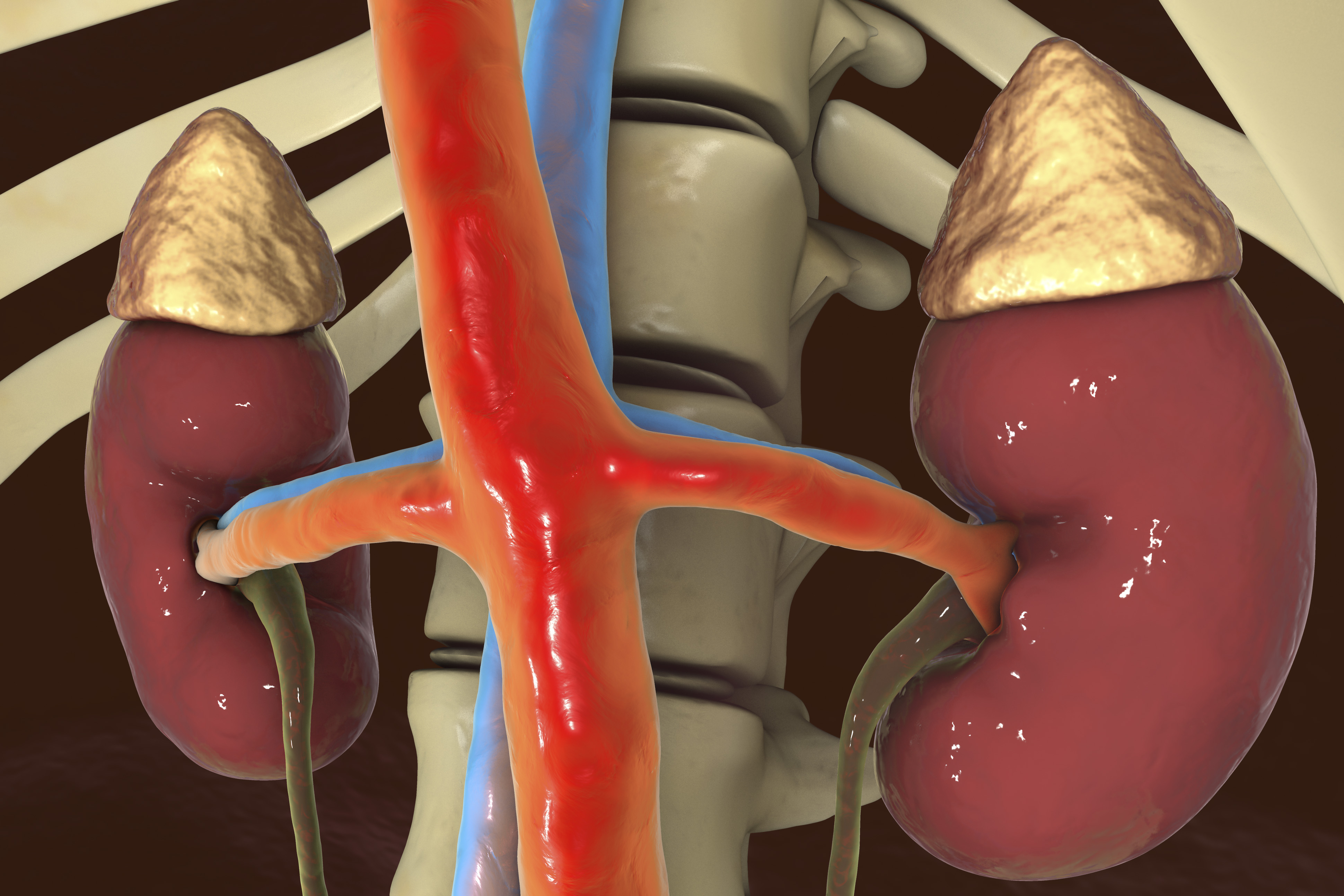
The ureters are small tubes that carry urine from each kidney to the bladder. A ureteral stone begins as a tiny grain of undissolved material located where urine collects in the kidney. When the urine flows out of the kidney, this grain of undissolved material is left behind. Over time, more undissolved material is deposited, and the stone becomes larger. Most stones enter the ureter when they are still small enough to move down into the bladder. From there, they pass out of the body with urination. However, some stones grow so large, that they become trapped in a narrow part of the ureter, causing pain and possibly blocking the flow of urine.
How are ureteral stones diagnosed?
Similar to kidney stones, ureteral stones may not be noticed until they begin to move or become lodged. Individuals with ureteral stones may experience several bothersome symptoms including:
- Extreme pain.
- Cramping.
- Blood in the urine (hematuria).
- Nausea.
- Vomiting.
- Fever.
- Chills.
- Painful urination.
- Frequent urination.
- A burning sensation when urinating.
Ureteral stone diagnosis
There are several diagnostic tools that can be used to detect ureteral stones. Stones are often found by x-ray or CT imaging. These diagnostic images provide urologists with valuable information about the stone’s size and location. If your doctor suspects a stone but is unable to make a diagnosis from a simple x-ray, they may scan the urinary system using a procedure called an intravenous pyelogram (IVP). Since an IVP requires preparation, it has been replaced in many hospitals by an abdominal/pelvic CT scan, an exceptionally accurate diagnostic tool that can detect almost all types of ureteral stones.
Urologists will also diagnose ureteral stones by a thorough review of symptoms, physical examination, urinalysis, and blood tests.
Treatment options
The appropriate treatment for ureteral stones largely depends on the size, position and number of stones. Small stones (0.2 inch or 4 mm in diameter) that are not causing infection, blockage or symptoms, have the ability to be flushed out by consuming large amounts of fluid. Pain can usually be treated with rest and analgesics (painkillers). Surgery is generally reserved for cases when conservative approaches have failed.
Surgery may be needed if:
- The stone will not pass based on its position or size.
- Individual is in extreme pain.
- Stone is blocking the flow of urine.
- The stone is associated with an ongoing urinary tract infection.
- The stone damages kidney tissue or causes constant bleeding.
- The stone continues to grow (as seen on follow-up x-rays).
- The stone is solitary.
Primary treatment options include Extracorporeal Shock Wave Lithotripsy (ESWL), Percutaneous Nephrolithotomy, and Ureteroscopy.
If you are having symptoms that may be associated with a kidney or ureteral stone, contact Urology Austin to schedule an appointment with one of our urologists.